Exploring the Inside of a Head
Some first-hand observations on the anatomy of a mammalian head and a mammalian brain.
If we wanted to explore the anatomy inside a person's head directly, how could we go about it? One way would be to buy an atlas of human anatomy, and study the pictures. But what if we want to study a real head ourselves? At least one science supply company sells sheep heads cut in half, allowing you to see inside. (For more details about this company, see my notes on dissection supplies. I also once found a boutique butcher shop that sold halves of pig's heads; perhaps you can find a similar place.)
A Sheep's Head
The is the first sheep head that I received from the science supply company:
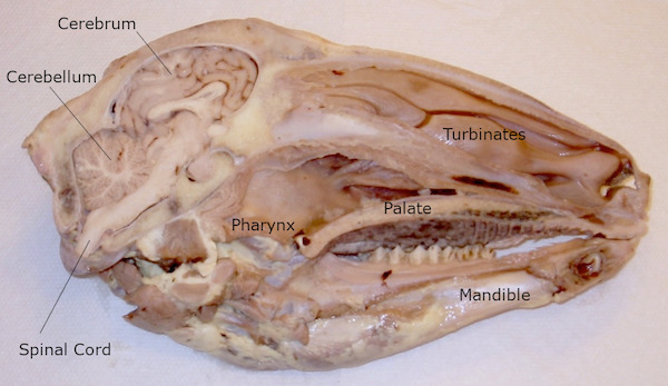
This specimen provided what I thought was a beautiful cross-section of the brain, while it was still in place inside a real head. You can notice in the photograph both of the main parts of the brain — the cerebrum and the cerebellum. And notice the proportions — the cerebellum and the cerebrum are nearly the same size, and the sheep's brain as a whole still occupies a small fraction of the space inside the sheep's head. A large part of the space inside a sheep's head is occupied by the nasal cavity. In a human head, about half of the head space is occupied by brain, and most of that is cerebrum. In a person, the cerebrum is much larger than the cerebellum, and the brain as a whole takes up a much larger proportion of the space.
Note, however, that the specimens vary considerably from one to the next. For example, this is the specimen I received the following year:
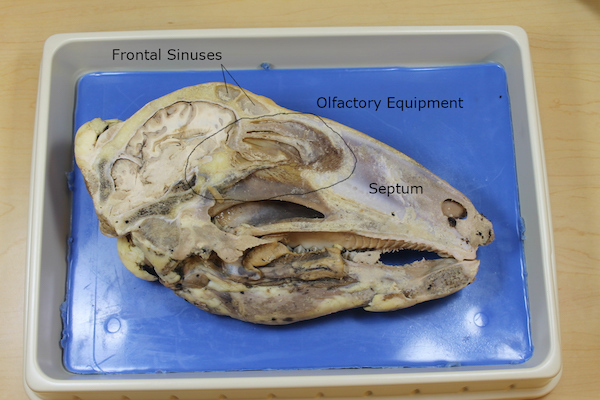
It is probably impossible to saw a head exactly through the nasal septum, so after slicing, one half of the head will contain the septum and the other half won't. The specimen I received the first year did not have the septum, allowing me to have a better view of the nasal passages, and the specimen I received the next year had the septum, blocking my view. On the other hand, the second specimen gave a clearer view of the olfactory apparatus. You can see in a rough way in the photo above how the brain grows through the base of the cranium to become the skin covering the complex structures at the top of the nose.
(What about the other side of the specimen, i.e. the outside of the head? I found the outside of the skinned head to be, shall we say, quite ugly. You could perhaps make some interesting observations about cheek muscles, and if you are ambitious perhaps you could cut around the eyeball to study the muscles that make it move. And it just occurred to me that by digging you might be able to trace the entire length of the optic nerve from the back of the eyeball to the optic chiasm at the base of the brain. But for the most part I just ignore the outside portion of the head. That is, until the kids beg to see it.)
A Sheep's Brain
Sheep's heads are relatively hard to find. But most science supply companies sell individual eyeballs, so that you can dissect one, and discover for yourself how an eyeball works. They also often sell brains.
I once found a specialty meat company that sold frozen brains. I meant to buy one and to see if I could thaw it and dissect it, and I regret that I never got around to it, because I have never found frozen brains since. However, preserved sheep brains from a science supply company are readily available, and are fairly inexpensive. There isn't much “dissecting” that you can do to a brain, but you can make some interesting observations about how it is connected to other things, and you can slice it down the middle and see a few things in the interior.
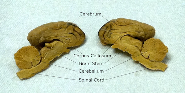
The top side of the brain is comparatively featureless, but you can clearly see the ridges and grooves (“gyri” and “sulci”) formed by the convolutions of the brain surface, and you can clearly see how the cerebrum is divided into two “hemispheres.”
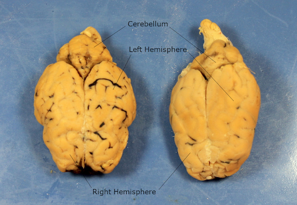
(Both of the brains are from sheep. I simply thought it might be interesting to compare two brains to see if there were any noteworthy variations from one individual to another.)
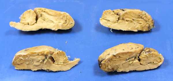
The bottom side of the brain is more complex, although the features are a little messier to discern. Perhaps the most noteworthy thing is the difference in color, and the shape of the whitish features.
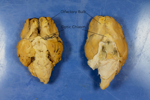
Notice that the brain itself is darker, and much of the core is more whitish. Perhaps we could call these materials “gray matter” and “white matter”? Gray matter makes up more of the “organ” of the brain itself, and white matter seems to be more for linking things together. In particular, notice two especially large features of white matter near the front of the brain, behind the sheep's face.
Immediately underneath the front portion of the cerebrum, in a position that places them directly above the nose, are a pair of white cables emerging from the core of the brain. You might think that the ends are swollen a little bit into “bulbs.” If you study an atlas of anatomy, you might discover that these bulbs send fibers through a perforated section of the floor of the cranium (the cribriform plate) and into a special patch of sensitive skin at the top of the nose. If you are lucky, you might be able to study this formation directly in a half of a sheep's head. These two cables are the olfactory nerves, and the bulbs are known as the olfactory bulbs. These two cables of white matter link the skin in your nose with your brain, and apparently help you to smell things. Maybe the skin “feels” the vapors somehow, and then tells the brain through the olfactory nerves?
Closer to the brain stem, you might notice a V-shaped structure, also made of white material. The tip of the V sits a little lower in the head, and a little farther back ... right behind the eyes. Can you guess what this is? If the brain still had the eyeballs attached, you would see that the two optic nerves connect to the tip of this V-shape, forming an X-shape in a living head. This X-shape is known as the optic chiasm, and it links the eyeballs to the brain. (You can find out what's at the other end of the optic nerve by dissecting an eyeball.)
These two pairs of large nerves — the olfactory and optic nerves — are actually two of twelve pairs of nerves that emerge from the base of the brain, collectively known as cranial nerves. The brain is connected to the entire body either through the spinal cord or through cranial nerves. Cranial nerves run mostly to the muscles and sense organs in the head while the spinal cord takes care of the rest of the body, but there is one cranial nerve (the vagus nerve) that runs to the internal organs in the torso. The olfactory nerves and the optic chiasm are probably the easiest to find, but you may be able to find more cranial nerves by carefully studying the base of the sheep's brain. A magnifying glass and a small probe might help.
After carefully examining the exterior of the brain, try carefully slicing the brain in half between the hemispheres. You will have to cut through something very tough, almost like a ligament holding the two halves together. That's the corpus callosum, and you may notice that it is made of white matter. You will also have to slice through the cerebellum, because the cerebellum doesn't have hemispheres.
I had one preserved specimen that I kept in fluid in a jar for years, and there were always tatters of fabric hanging off of it. With dry specimens, you might see more clearly that all of the structures of the brain are carefully wrapped in layers of protective fabric. These are the meninges.