The Thoracic Organs of a Sheep
The heart and lungs form a tightly integrated package working together to energize the body, and you can study how this organ bundle works by dissecting “sheep pluck”.
I generally recommend dissecting offal over dissecting preserved organs. Fresh organs are cheaper and the colors and textures are more realistic. They are also less smelly, unless you keep them for too long. Unfortunately, there is one major organ that you can't buy for food in the United States: lungs. Selling lungs as offal is illegal, even though lungs are commonly eaten in other countries. (I've always wanted to try authentic haggis, and I'm annoyed that you can't obtain it in the United States.) So if you live in the United States and you want to dissect fresh lungs, I'm afraid you're out of luck.
You can still buy preserved lungs, however. They are normally sold as part of the entire bundle of thoracic organs (normally from sheep). The heart and lungs are actually tied very tightly together into a single unified organ package, and it is much easier to “pluck” this bundle from the ribcage than it is to separate the individual organs from each other. (To purchase sheep pluck, I recommend Carolina Biological Supply.)
The reason these particular organs are tied so tightly together, and nestled together as a bundle inside the protective ribcage, is because they need to collaborate, both pumping constantly and working together to keep your body continuously energized with fresh warm blood. Perhaps we could think of this thoracic organ bundle as the “engine” of a mammal. It's not a perfect analogy — the organs that actually make motion happen are the muscles. But these pumping organs in the chest run constantly to keep the rest of the body energized and warm. They “rev up” when you are being extra active, and they can slow down to an idle when you are resting, but they can never stop as long as you live. So I think it's a very helpful analogy. (Feel free to discuss the pros and cons of the engine analogy explicitly with your kids. It's good “thinking practice”.)
So dissecting “sheep pluck” is more than just a way to explore what lungs are like. It's a good way to explore how they interact with the heart, and to explore the entire “engine” that you have in your chest.
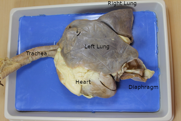
(This particular specimen had the right lung deformed backwards, as if it were coming out the animal's right shoulder, as a consequence of the preservation process. And whoever prepared it carelessly nicked the heart with a stray cut. If the trachea included the layrnx on top, you could study how a voice box works, but my speciment never included a larynx.)
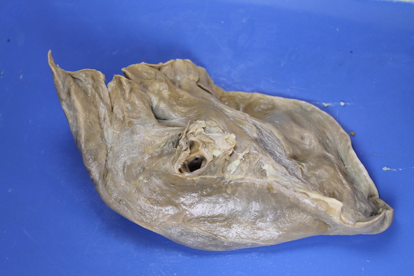
One of the first things you might notice is that the lungs are not sacks or bags. They are much more sponge-like than sac-like, and they are quite dense and heavy. (I would usually try to demonstrate that these sponges could be inflated by blowing into the trachea. If you can do this successfully, it is a very memorable demonstration. However, your ability to do this will depend on the quality of the specimen. If the trachea is damaged, your breath will pass out the openings instead of into the lungs. You also have to be willing to use the end of the sheep's trachea like a mouthpiece. You may prefer to place something over the end of the trachea before you place your lips against it, but it has to allow your breath through the middle easily while still sealing around the edges. As an alternative, sometimes after removing a lung you can manage to inflate a small portion of a lung by pressing a syringe full of air against one of the bronchi in the hilum. You might even be able to demonstrate that different bronchi go to different lobes of the lung by trying different bronchi.)
Another thing you might notice is that the lungs have a few separate “flaps” or “lobes”. Human beings typically have three major lobes in the right lung, and two in the left, and you may find it interesting to see if you can identify and count the lobes in your sheep specimen. You might also notice all of the sticky fabric that stitches and sticks together the various lobes and the various organs, helping to form a single “organ bundle”:
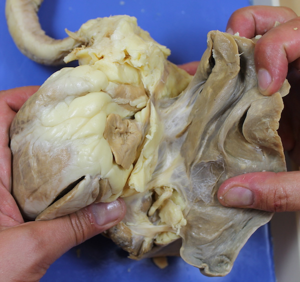
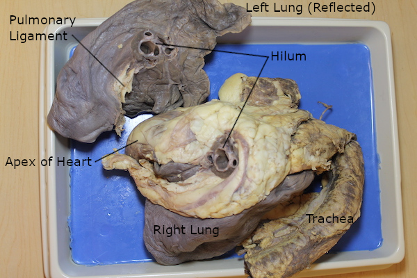
If you've ever dissected a heart, you may have noticed the “tube tangle” at the top. This knot of tubes ties the heart very tightly together with the lungs. If you tear apart the connective cobwebs, and try to find where the tubes run from the tube tangle into the lungs, you find that everything that enters or leaves a lung passes through a single “doorway”. We call this the hilum of the lung. A branch from the trachea and several tubes from the heart's “tube tangle” run through this doorway into each lung. (If your trachea is very short, perhaps you can pass a long probe of some kind down the trachea and make the end enter one of the lungs.)
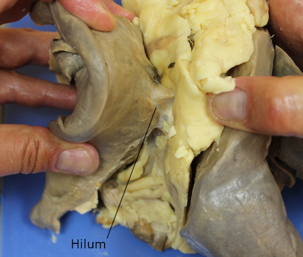
If you slice through this hilum, you can separate a lung from the rest of the package, and study the tubes that pass through the doorway. (You will probably also have to cut an especially thick ligament extending downwards from the hilum, sewing the lung tightly to the rest of the bundle. That's the pulmonary ligament.) If you inspect the tubes carefully, you may be able to identify three different kinds of tubes. One kind has solid, cartilaginous walls, and if you pass a probe of some kind into the bundle-side of one of these tubes, you should find that it enters the trachea. These tubes are called bronchi, and they are apparently for carrying air. (Why do you suppose all of the air-tubes are made of cartilage?) The rest of the tubes have fabric walls, and you may find upon careful inspection that some have thicker walls, as if they have a small amount of muscle surrounding them, and the others have merely a thin sheet of sealant. If you try to find out where these tubes go in the organ bundle, you may be able to determine that they lead to the heart. These two types of fabric tubes are presumably for carrying blood to and from the heart. (By the way, “reflected” means folded or flipped over. If you study the pictures in an atlas of anatomy, you'll see this term often.)
If you study the contours of the lung carefully, you may be able to find that they correspond to other neighboring organs. Soft spongy organs like the lungs don't really need to have any specific shape, so they can just fill whatever space is available, and leave room wherever needed for their neighbors. With preserved organs, however, this can be difficult. The handling and the preservative process can harden the sponges into unnatural and unrealistic shapes, so you may or may not be able to learn anything by studying the shape. (This is also why the right lung in the first picture above is sticking out at a strange angle.)